A Deeper Look at Compression Fractures
As we age, the continual process of replacing bone tissue slows down causing your bones to become thinner and weaker. If this becomes severe enough, you could develop osteoporosis, which is a condition that significantly raises the risk of fractures. Simple daily activities like standing, walking or sneezing could result in fractures among people with severe osteoporosis.
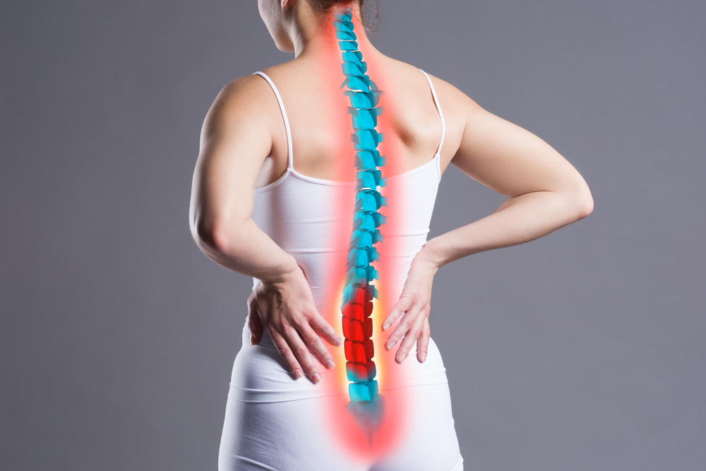
In certain people with osteoporosis, the spinal vertebrae may become so weakened that they cannot support the spinal column. This may produce spinal compression fractures. Spinal compression fractures are usually accompanied by a deep ache in the area of the fracture along with sensitivity to touch. You may also experience pain while breathing or moving.
What Is Osteoporosis?
In the United States, there are almost 44 million people with osteoporosis and another 3 million with low bone mass. About half of all Americans aged 50 or older live with osteoporosis, and one in two Asian or Caucasian women over 50 will break a bone due to osteoporosis.
This disease causes bones to break by weakening the internal structure of bones. In healthy bones, there are numerous hollow spaces surrounded by thick bony walls, but in people with osteoporosis, the spaces grow much bigger as the bony structures thin.
There are many causes of osteoporosis, but, by far the most common, is aging. Throughout your life, your body removes old bone and replaces it with new bone. However, after age 30, your body begins to break down more bone than it can replace. Even among healthy older people, therefore, there is an elevated risk of fractures.
Another contributing factor for osteoporosis is menopause. The hormonal changes that accompany menopause among women at 45 or older causes bone loss to accelerate. However, following menopause, this pace of bone loss slows.
There are other risk factors for osteoporosis, including
- Being Caucasian or Asian
- Poor diet
- Sedentary lifestyle
- Tobacco use
- Low body weight
- Female
- Family history of osteoporosis
If you are diagnosed with osteoporosis, there are a few treatment options. First of all, your doctor will probably recommend some lifestyle changes that include a diet high in calcium and vitamin D, as well as more weight-bearing exercise. If necessary, your doctor may also recommend drugs called bisphosphonates. Hormone therapies—testosterone for men and estrogen for women—may be helpful to certain patients. If you smoke, your doctor will strongly encourage you to stop because of the damage it does to your bones.
Spinal Compression Fractures
Although osteoporosis is the most common cause of spinal compression fractures, it is not the only one. Of course, you may experience a spinal compression fracture if you are involved in a collision. Spinal tumors may also produce spinal compression fractures by eroding the bone structure.
To understand what happens when a spinal compression fracture occurs, you must first learn about the spine. There are two primary parts to the spine: vertebral arch and vertebral body. The vertebral arch is the roof the spine and can be felt by placing your hand on the back. The vertebral body is the cylindrical part that encases the spinal cord.
In most spinal compression fractures, the vertebral body collapses while the arch remains intact. That is why many people with spinal compression fractures exhibit kyphosis in which the upper back is curved forward.
Although there may be apparent symptoms of a spinal compression fracture that include pain, limb weakness or numbness, or loss of height, some people may not realize that a spinal compression fracture has occurred. In many cases, the fracture may gradually appear and only produce mild symptoms that are easily dismissed as a dull ache or other minor feature.
Diagnosing a Compression Fracture
In order to diagnose a spinal compression fracture, your doctor should begin by taking your medical history and performing a thorough physical examination. If there is reason to believe that a spinal compression fracture has occurred, then your doctor may order an x-ray which will image the bones of the spine.
In some cases, it may be necessary to obtain more detailed imagery of the spinal region, so an MRI or CT scan may be performed. Both of these kinds of scans can provide much more information about the spinal vertebrae as well as surrounding soft tissue like muscles, fat and nerves. If further analysis of the bone is required, then a nuclear bone scan in which a mildly radioactive dye is injected into the bones is performed.
Treating Spinal Compression Fractures
In most cases, spinal compression fractures can be treated conservatively. It typically takes about three months for a spinal compression fracture to fully heal. During this period, your doctor may recommend mild pain relievers or more potent prescription medications depending on the severity of the pain symptoms. You may also be asked to wear a back brace which should help straighten your spine and relieve pressure on the fractured vertebrae.
If the primary cause of the spinal compression fracture was osteoporosis, then your doctor may implement a treatment plan to help strengthen your bones. This may include weight-bearing exercises, bisphosphonates and a diet rich in calcium and vitamin D.
If your spine is unstable due to compression fractures, then a surgical procedure may be necessary.
- Vertebroplasty—during this procedure, the surgeon will use guided imagery to inject a cement compound into the fracture to stabilize the bone. After an hour in which the cement hardens, you may be allowed to return home.
- Kyphoplasty—a kyphoplasty is very similar to a vertebroplasty, but this procedure requires inflating a small balloon in the vertebra first to create a space for the bone cement.
- Spinal fusion surgery—if the spinal compression fracture is producing pain due to an overly mobile vertebra, then a spinal fusion procedure may be necessary. During this procedure, some bone is harvested, usually from your hip, and then grafted into the space between two vertebrae. Your body will then use the bone graft as scaffolding to grow new bone that fuses and immobilizes the two vertebrae.
Article written by: Dr. Robert Moghim – CEO/Founder Colorado Pain Care
M.D. Disclaimer: The views expressed in this article are the personal views of Robert Moghim, M.D. and do not necessarily represent and are not intended to represent the views of the company or its employees. The information contained in this article does not constitute medical advice, nor does reading or accessing this information create a patient-provider relationship. Comments that you post will be shared with all visitors to this page. The comment feature is not governed by HIPAA and you should not post any of your private health information.

Colorado Pain Care treats each patient with the same care we would want for our own family. Founded on the promise of H.O.P.E., we provide honest, objective, personalized, and empathetic care from the area’s top physicians and providers.

